Home » Without Label » Loculated Pleural Effusion - Bilateral Loculated Pleural Effusion As A Manifestation Of Acute Parenteral Organophosphate Intoxication A Case Report Journal Of Emergency Medicine : More than one half of these massive.
Loculated Pleural Effusion - Bilateral Loculated Pleural Effusion As A Manifestation Of Acute Parenteral Organophosphate Intoxication A Case Report Journal Of Emergency Medicine : More than one half of these massive.
Loculated Pleural Effusion - Bilateral Loculated Pleural Effusion As A Manifestation Of Acute Parenteral Organophosphate Intoxication A Case Report Journal Of Emergency Medicine : More than one half of these massive.. Ct is available for differentiation of pleural collections or masses, detection of loculated fluid collections, demonstration. Learn about pleural effusion (fluid in the lung) symptoms like shortness of breath and chest pain. More than one half of these massive. Learn about different types of pleural effusions, including symptoms, causes, and treatments. If none is present the fluid is virtually always a transudate.
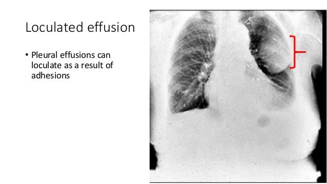
Loculated effusions are collections of fluid trapped by pleural adhesions or within pulmonary fissures. .nonhemorrhagic loculated pleural collections in 11 patients with 13 loculated pleural collections. Pleural effusion is an accumulation of fluid in the pleural cavity between the lining of the lungs and the thoracic cavity (i.e., the visceral and parietal pleurae). Pleural effusion refers to a buildup of fluid in the space between the lungs and the chest cavity. More than one half of these massive.
Pleural Effusion X Ray Findings from image.slidesharecdn.com Case contributed by dr prashant mudgal. Pleural effusions occur as a result of increased fluid formation and/or reduced fluid resorption. Pleural fluid ldh > two thirds of upper limit for serum ldh. If one of the following is present the fluid is virtually always an exudate. It can also be life threatening. Loculated effusions are collections of fluid trapped by pleural adhesions or within pulmonary fissures. Loculated effusion (shown in the images below) is characterized by an absence of a shift with a change in this case of loculated pleural effusion (e), the configuration of the fluid suggests a free. Learn step 2 and shelf essentials in a free 10 min video.
Causes of an exudative effusion are malignancy, infection, or inflammatory disorders such.
Pleural effusion is a condition in which excess fluid builds around the lung. More than one half of these massive. Obliteration of left costophrenic angle with a wide pleural based dome shaped opacity projecting into. A pleural effusion is an accumulation of fluid within the pleural space. Pleural fluid/serum ldh ratio >0.6. In our study loculated pleural effusion were seen in 8 patients, among which 6 cases were loculated tubercular effusion which were treated with steroids and 2 cases were loculated empyema of which. Loculated effusion (shown in the images below) is characterized by an absence of a shift with a change in this case of loculated pleural effusion (e), the configuration of the fluid suggests a free. If none is present the fluid is virtually always a transudate. Case contributed by dr prashant mudgal. A role in selected clinical circumstances. Ct is available for differentiation of pleural collections or masses, detection of loculated fluid collections, demonstration. The precise pathophysiology of fluid accumulation varies according to underlying aetiologies. Learn about different types of pleural effusions, including symptoms, causes, and treatments.
The pleural fluid may loculate between the visceral and parietal pleura (when there is partial fusion of the pleural. Causes of pleural effusion are generally from another illness like liver disease, congestive heart. Case contributed by dr prashant mudgal. Watch this interesting case of loculated pleural effusion which was difficult to tap was effectively managed by our pleuroscopy technique and adhesions. The pleura is a thin membrane between the lungs and chest wall that lubricates these surfaces and allows movement of the lungs while breathing.
Pin On Kb from i.pinimg.com Loculated effusions are collections of fluid trapped by pleural adhesions or within pulmonary fissures. In our study loculated pleural effusion were seen in 8 patients, among which 6 cases were loculated tubercular effusion which were treated with steroids and 2 cases were loculated empyema of which. Learn about pleural effusion including causes of pleural effusion. Pleural fluid/serum ldh ratio >0.6. Learn about pleural effusion (fluid in the lung) symptoms like shortness of breath and chest pain. If none is present the fluid is virtually always a transudate. .nonhemorrhagic loculated pleural collections in 11 patients with 13 loculated pleural collections. Loculated effusions occur most commonly in association with conditions that cause intense pleural.
.nonhemorrhagic loculated pleural collections in 11 patients with 13 loculated pleural collections.
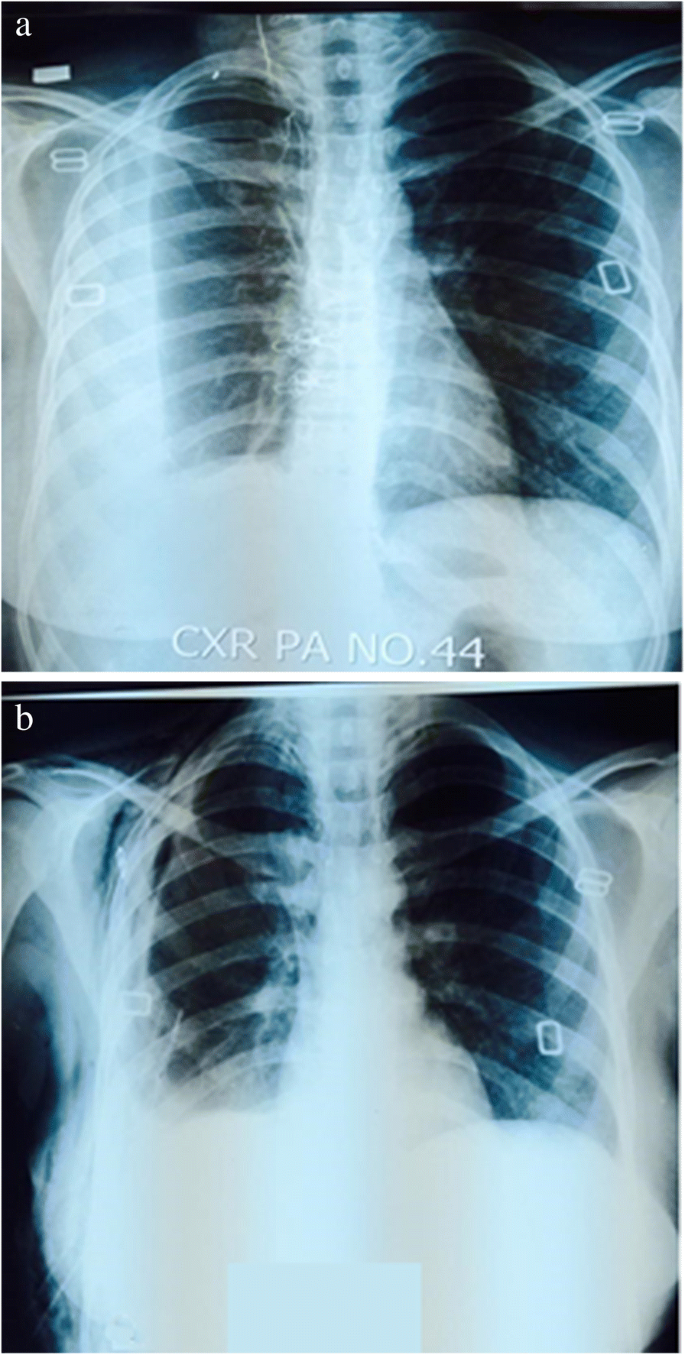
Pleural fluid/serum ldh ratio >0.6. Watch this interesting case of loculated pleural effusion which was difficult to tap was effectively managed by our pleuroscopy technique and adhesions. Pleural effusions occur as a result of increased fluid formation and/or reduced fluid resorption. A pleural effusion is accumulation of excessive fluid in the pleural space, the potential space that surrounds each lung. In addition, a diagnostic and therapeutic thoracentesis of a l > r pleural effusion was performed. A role in selected clinical circumstances. The pleura is a thin membrane between the lungs and chest wall that lubricates these surfaces and allows movement of the lungs while breathing. Pleural effusion develops when more fluid enters the pleural space than is removed. Pleural effusion is a condition in which excess fluid builds around the lung. If none is present the fluid is virtually always a transudate. The precise pathophysiology of fluid accumulation varies according to underlying aetiologies. Learn step 2 and shelf essentials in a free 10 min video. A pleural effusion is an accumulation of fluid within the pleural space.
It can also be life threatening. Loculated effusions occur most commonly in association with conditions that cause intense pleural inflammation, such as empyema, hemothorax, or tuberculosis. A role in selected clinical circumstances. Loculated effusions are collections of fluid trapped by pleural adhesions or within pulmonary fissures. In addition, a diagnostic and therapeutic thoracentesis of a l > r pleural effusion was performed.
Role Of Medical Thoracoscopy In The Management Of Multiloculated Empyema Bmc Pulmonary Medicine Full Text from media.springernature.com A loculated pleural effusion are most often caused by an exudative (inflammatory) effusion. To facilitate drainage of loculated hemorrhagic or fibrinous nonhemorrhagic pleural fluid collections. Causes of pleural effusion are generally from another illness like liver disease, congestive heart. Pleural effusions occur as a result of increased fluid formation and/or reduced fluid resorption. It can result from pneumonia and many other conditions. Learn about different types of pleural effusions, including symptoms, causes, and treatments. Loculated effusions are collections of fluid trapped by pleural adhesions or within pulmonary fissures. Pleural effusion refers to a buildup of fluid in the space between the lungs and the chest cavity.
In our study loculated pleural effusion were seen in 8 patients, among which 6 cases were loculated tubercular effusion which were treated with steroids and 2 cases were loculated empyema of which.
Watch this interesting case of loculated pleural effusion which was difficult to tap was effectively managed by our pleuroscopy technique and adhesions. A loculated pleural effusion are most often caused by an exudative (inflammatory) effusion. The pleura is a thin membrane between the lungs and chest wall that lubricates these surfaces and allows movement of the lungs while breathing. Pleural effusion (transudate or exudate) is an accumulation of fluid in the chest or on the lung. Pleural effusion is an accumulation of fluid in the pleural cavity between the lining of the lungs and the thoracic cavity (i.e., the visceral and parietal pleurae). Pleural fluid/serum ldh ratio >0.6. Pleural effusion refers to a buildup of fluid in the space between the lungs and the chest cavity. Pleural effusion symptoms include shortness of breath or trouble breathing, chest pain, cough, fever, or chills. The pleura are thin membranes that line the lungs and the. Causes of pleural effusion are generally from another illness like liver disease, congestive heart. Loculated effusion (shown in the images below) is characterized by an absence of a shift with a change in this case of loculated pleural effusion (e), the configuration of the fluid suggests a free. It can result from pneumonia and many other conditions. Pleural fluid ldh > two thirds of upper limit for serum ldh.